
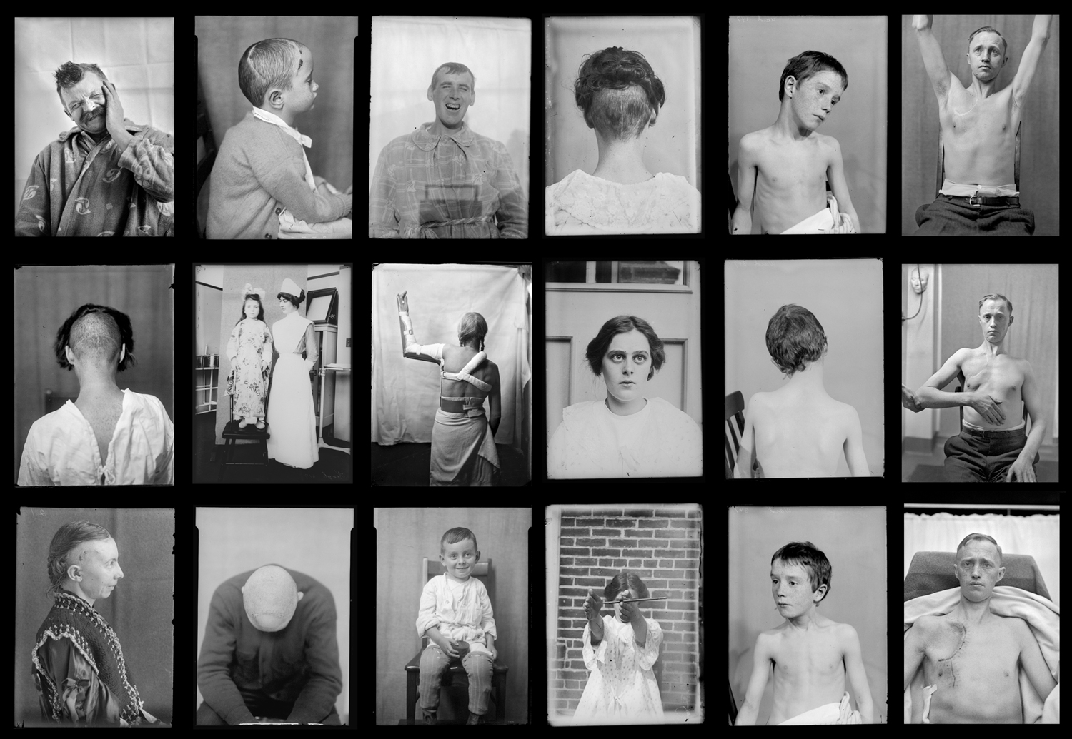
Some of the approximately 10,000 patient images from the Cushing Tumor Registry.
United States, especially at Johns Hopkins Hospital in Baltimore, Maryland, and Peter Bent Brigham Hospital in Boston, Massachusetts
Harvey Cushing; Terry Dagradi; others
The photographic materials in this unique historical collection consist of approximately 10,000 to 15,000 glass and film (some, probable cellulose nitrate) negatives, and several hundred lantern slides. The photographs, taken from 1905 to 1932, provide records of patients, surgical techniques, examples of patient tumors, and, in some cases, the brains of deceased patients from autopsy. Also included are portraits of patients, pre- and post-operative images, autopsy photographs, gross specimen photographs, microscope histology slides, diagnostic optical charts, and patient case composites. Only a fraction of the photographs have been viewed. Eventually all the photographic materials in the collection will be digitized and available through a searchable database.
The collection contains the records of Dr. Harvey Cushing (Yale 1891), noted pioneer and world-renowned teacher of brain surgery. Most were taken at the Peter Bent Brigham Hospital in Boston, Massachusetts, and at the Johns Hopkins Hospital in Baltimore, Maryland. The photographs are connected to the collection’s approximately 640 jars of preserved brains and specimens, and to actual patient records.
